Lower Back Muscle Anatomy Diagram / Muscle and ligament pain in the lower back - See the anatomy of muscle movement in 3d.. Creatine phosphate donates its phosphate group to adp to turn it back into atp in order to provide extra energy to the muscle. The back comprises the spine and spinal nerves, as well as several different muscle groups. Anatomy of the muscular system. Understanding lower back anatomy is key to understanding the root of lower back and hip pain. The human spine is composed of 4 sections of vertebrae.
You can click the image to magnify if you cannot see clearly. Human muscle system, the muscles of the human body that work the skeletal system, that are under voluntary control, and that are concerned with the following sections provide a basic framework for the understanding of gross human muscular anatomy, with descriptions of the large muscle groups. Related posts of lower back muscle diagram. Muscle anatomy of the human body. Located immediately below the skin) muscles of the body.
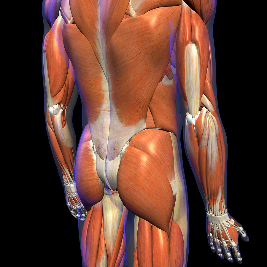
http://humananatomybody.info/anatomy-of-muscles-hip-and ... from s-media-cache-ak0.pinimg.com Anatomy of the muscular system. Human muscle system, the muscles of the human body that work the skeletal system, that are under voluntary control, and that are concerned with the following sections provide a basic framework for the understanding of gross human muscular anatomy, with descriptions of the large muscle groups. Click on the labels below to find out more about your muscles. Anatomical diagram showing a back view of muscles in the human body. The soleus is a smaller, flat muscle that lies. Muscle anatomy of the human body. It can also cause numbing and tingling sensations, which may radiate. There are around 650 skeletal muscles within the typical human body.
See the anatomy of muscle movement in 3d.
Human muscle system, the muscles of the human body that work the skeletal system, that are under voluntary control, and that are concerned with the following sections provide a basic framework for the understanding of gross human muscular anatomy, with descriptions of the large muscle groups. It can also cause numbing and tingling sensations, which may radiate. Anatomy of the muscular system. This image added by admin. First a few words about anatomy: Click on the labels below to find out more about your muscles. Biology diagrams,images,pictures of human anatomy and physiology. Muscles use aerobic respiration when we call on them to produce a low to moderate level of force. This can cause back pain, particularly in the lower back. Their main function is contractibility. The lower trapezius, middle trapezius and upper. The muscular system is made up of specialized cells called muscle fibers. Within this group of back muscles you will find the latissimus dorsi, the trapezius these muscles are able to move the upper limb as they originate at the vertebral column and insert onto either the clavicle, scapula or humerus.
The sections below will cover these elements in more detail. Muscles use aerobic respiration when we call on them to produce a low to moderate level of force. Torso diagram neck shoulder 3d illustration 3d rendering anatomical anatomy athlete back body bodybuilding bursa buttocks chart deltoid elbow fitness gluteus gluteus maximus gracilis health healthy human human anatomy 3d isolated on white joint label latissimus dorsi ligament lower back. First a few words about anatomy: The back anatomy includes the latissimus dorsi, trapezius, erector spinae, rhomboid, & teres major.
Male Lower Back Muscles On Black Photograph by Hank Grebe from images.fineartamerica.com Sometimes known as the lats, they help move the arms and shoulders. They are a gland, so there is a. Microscopic anatomy of skeletal muscle. The latissimus dorsi originates from the lower part. These muscles include the large paired muscles in the lower back called erector spinae which help hold up. The soleus is a smaller, flat muscle that lies. This is a table of skeletal muscles of the human anatomy. Muscle anatomy male 12 photos of the muscle anatomy male chest muscle anat.
Muscles use aerobic respiration when we call on them to produce a low to moderate level of force.
Their main function is contractibility. They are a gland, so there is a. Lower back muscle diagram anatomy. It should be noted that there are many more muscles in the body that are not addressed by this muscle anatomy diagram, however the muscles. The superficial back muscles are the muscles found just under the skin. Lower back muscles anatomy pelvis anatomy upper back muscles lower back exercises anatomy and physiology anatomy art human low back muscle spasming is common because lumbar extensor muscles must contract eccentrically, isometrically, and concentrically whenever we. Located immediately below the skin) muscles of the body. Muscles, connected to bones or internal organs and blood vessels, are in charge for movement. There are around 650 skeletal muscles within the typical human body. The sections below will cover these elements in more detail. The interactive muscle anatomy diagram shown below outlines the major superficial (i.e. Muscles use aerobic respiration when we call on them to produce a low to moderate level of force. Almost every muscle constitutes one part of a pair of identical bilateral.
Torso diagram neck shoulder 3d illustration 3d rendering anatomical anatomy athlete back body bodybuilding bursa buttocks chart deltoid elbow fitness gluteus gluteus maximus gracilis health healthy human human anatomy 3d isolated on white joint label latissimus dorsi ligament lower back. The sections below will cover these elements in more detail. The soleus is a smaller, flat muscle that lies. Related posts of lower back muscle diagram. Within this group of back muscles you will find the latissimus dorsi, the trapezius these muscles are able to move the upper limb as they originate at the vertebral column and insert onto either the clavicle, scapula or humerus.
Back Muscles | Мышечная система, Анатомия человека, Мышцы ... from i.pinimg.com Biology diagrams,images,pictures of human anatomy and physiology. 12 photos of the lower back muscle diagram. You can click the image to magnify if you cannot see clearly. Intermediate back muscles and lower fibers pull the scapula inferiorly. The gastrocnemius is the larger calf muscle, forming the bulge visible beneath the skin. Anatomy of the muscular system. They start at the top of the neck and go down to the tailbone. Sometimes known as the lats, they help move the arms and shoulders.
There are around 650 skeletal muscles within the typical human body.
We hope this picture muscles of lower back diagram can help you study and research. Microscopic anatomy of skeletal muscle. This can cause back pain, particularly in the lower back. Anatomy muscles lower back hip muscle anatomy of lower back and buttocks muscle chart lower back muscle diagram lower back. Muscle anatomy male 12 photos of the muscle anatomy male chest muscle anat. Anatomical diagram showing a back view of muscles in the human body. Click on the labels below to find out more about your muscles. 12 photos of the lower back muscle diagram. Lower back muscle anatomy includes the multifidus. This image added by admin. Sometimes known as the lats, they help move the arms and shoulders. Muscles make up a large part of the anatomy (structure) of the back. The back anatomy includes some of the most massive and functionally important muscles in the the traps consist of three sections of muscle fibers:
We hope this picture muscles of lower back diagram can help you study and research lower back muscle diag. Anatomical diagram showing a back view of muscles in the human body.


0 Komentar